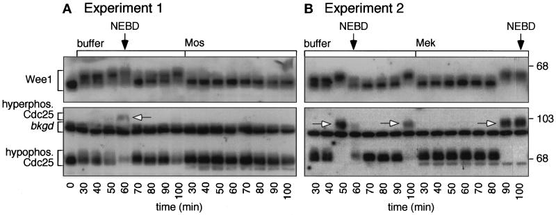
Figure 7.
Cdc25 and Wee1 phosphorylation in MAP kinase-activated extracts. Cycling extracts were prepared and supplemented with sperm chromatin and Mos, Mek, or buffer. Samples were removed every 10 min for analysis. (A and B) Immunoblots of Cdc25 and Wee1. Top, autoradiograms show the periodic accumulation of hyperphosphorylated Wee1 in the buffer-treated samples (left), with the most highly shifted forms peaking at nuclear envelope breakdown (NEBD). In addition, at mitosis the intensity of the hypophosphorylated Cdc25 band is reduced (lower), and some hyperphosphorylated Cdc25 is detectable (arrows). Some hyperphosphorylated Cdc25 may be obscured by the indicated background band. There is no change in electrophoretic mobility of either Wee1 or Cdc25 in the Mos- or Mek-treated extracts (right); both proteins remain in their hypophosphorylated interphase forms.

