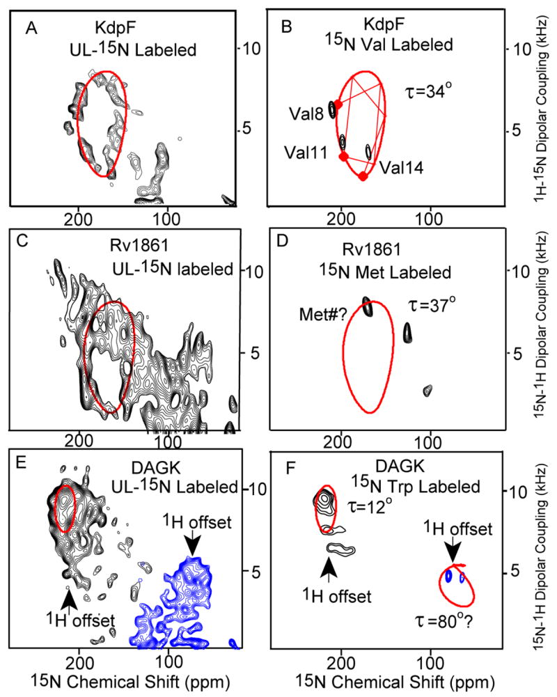
Figure 2.

PISEMA spectra of KdpF (A&B), Rv1861 (C&D) and DAGK (E & F) of uniformly 15N-labeled (A,C&E) and amino acid specific labeled protein (B,D&F) expressed in E. coli and reconstituted into a mixture of lipids – dimyristoylphosphatidylcholine (DMPC) and phosphatidyl-glycerol (DMPG) in a 4:1 molar ratio. The samples were aligned between glass slides (see supplemental materials). Spectra were obtained with NHMFL low-E probes at 600 MHz except for the 15N UL KdpF spectrum which was obtained in the UWB 900 MHz magnet. The low-electric field feature was essential for this spectroscopy. 0.8 ms cross polarization contact time, an acquisition time of 4 ms during with SPINAL9 decoupling was applied, and a recycle delay of 6 s were used. To avoid the limitations of the 1H bandwidth the spectra of DAGK were obtained in two halves with different offset frequencies. Spectra acquisition time varies from 6 hours to 3 days. The PISA wheels were calculated using motionally averaged dipolar (ν|| = 10.375 kHz) and chemical shift tensors (σ11 = 57; σ22 = 81; σ33 = 228 ppm)10,11.
