
Figure 8. S783 phosphorylation promotes neuronal survival after anoxia insult.
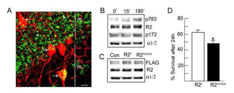
A. Analyzing the subcellular sites of S783 phosphorylation in the hippocampus using confocal microscopy. Hippocampal sections from MCAO animals were stained with antibodies against S783 and parvalbumin, a marker for subsets of interneurons, with secondary antibodies FITC and rhodamine respectively. Arrows indicate parvalbumin positive interneurons, white arrowheads represent parvalbumin negative interneurons, PL = pyramidal cell layer, RL = Stratum radiatum, scale bar = 10 μm. B. Anoxia increases AMPK activity and S783 phosphorylation. 12-14 DIV hippocampal neurons were given a 5 min anoxic insult, re-fed and incubated at 37°C for either 15 or 180 min as indicated. Neurons were then lysed and immunoblotted with antibodies against p783, GABABR2 (R2), p173 and AMPK α1/2 subunits (n=4-5). C. Expression of recombinant GABAB receptors in hippocampal neurons. 10-14 DIV hippocampal neurons expressing either GABABR2F, or GABABR2FS783A subunits were lysed and immunoblotted with FLAG antibodies. D. Mutation of S783 in GABABR2 decreases neuronal survival 24 hr after anoxia. Neurons were stained with trypan blue and viability was calculated as a percentage of unstained cells (viable cells) related to the total number of cells counted viable cells plus nonviable cells). At least 300 cells were counted on each coverslip. * = significantly different from control (p<0.01; students-t test, n=3)