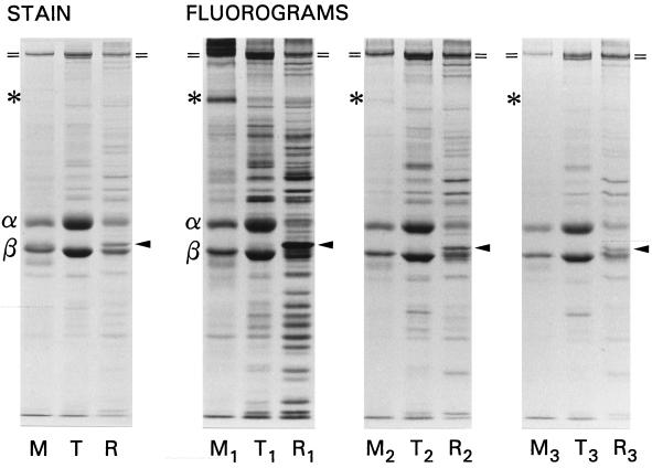
Figure 3.
SDS-PAGE and 3H-fluorographic analysis of ciliary proteins synthesized during one regeneration and replaced with unlabeled proteins in two successive regenerations. The detergent-solubilized membrane/matrix (M), thermally solubilized tubulin (T), and stable ninefold remnant (R) were loaded stoichiometrically; the subscripts denote the first, second, and third regenerations. A prominently labeled membrane protein is designated with an asterisk; the positions of the barely resolved dynein heavy chains are marked (=); the α and β tubulin chains are so designated; the solid arrowheads mark tektin A.

