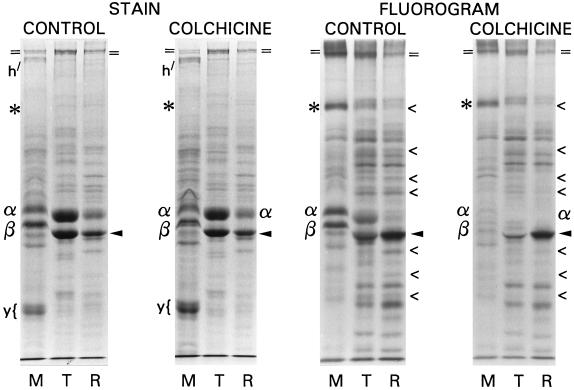
Figure 5.
SDS-PAGE and 3H-fluorographic analysis of ciliary protein fractions from control and 1 mM colchicine-treated midgastrula stage embryos. Left panel: Coomassie blue-stained gel; right panel: fluorogram of the same gel. Tubulin synthesis is inhibited by colchicine, but protein distribution and labeling are similar under both conditions. The retarded migration in the M lanes is due to the presence of hyalin, normally removed when embryos are predeciliated during culture, which these intentionally were not. Designations as in Figure 3; “<” denotes proteins whose labeling, but not amount, is diminished by colchicine. Nonciliary, nonlabeled proteins originating from high-salt solubilization of the hyaline layer during deciliation are noted: h, hyalin; y, a yolk granule component.

