
Figure 3. Immunocytochemistry and Western blot analysis of γH2AX expression.
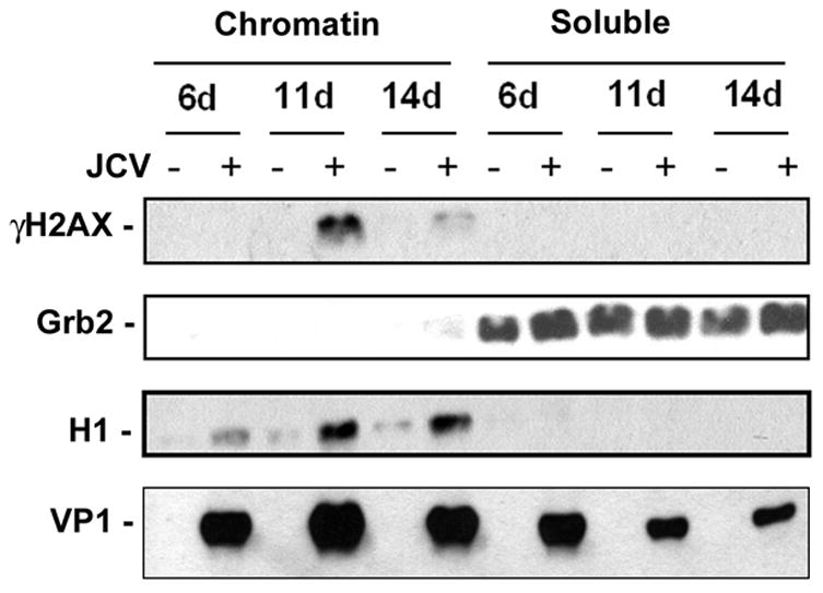
Mad-1/SVEdelta JCV-infected astrocytes and uninfected controls were harvested at 6, 11 and 14 days post-infection and fractionated into chromatin-associated and soluble components as described in Materials and Methods. γH2AX was measured by Western blot. H1 and Grb2 were used as fractionation and loading controls. Infection was verified by Western blot to VP1.