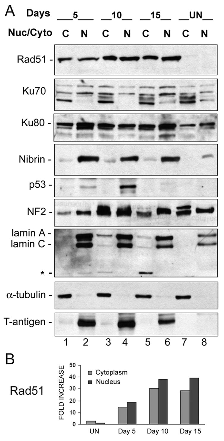
Figure 4. Expression of DNA repair proteins in the cytoplasmic and nuclear subcellular compartments.

A. Astrocytes infected with Mad-1/SVEdelta JCV for 5, 10 and 15 days and uninfected controls were subject to subcellular fractionation and cytoplasmic (C) and nuclear (N) proteins were harvested. These were subject to Western blot analysis for DNA repair proteins. To assess the purity of the fractions, α-tubulin was used as a marker for cytoplasm and lamin A/C as a marker for the nucleus. The asterisk indicates a band corresponding to the cleaved form of lamin (28 kDa). T-antigen was used as a marker for infection. Ku70 runs as multiple bands, which may be due to phosphorylation. B. Quantitation of the differences in Rad51 levels in Panel A was performed by densitometric analysis of the X-ray film scanned on a Molecular Imager FX with the Quantity One Program (Bio-Rad, Hercules CA). The results were normalized relative to expression of Rad51 in the nuclei of uninfected cells and are presented as a histogram.
