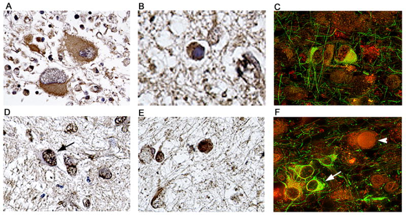
Figure 9. Detection and cellular localization of DNA repair enzymes Ku70 and Ku80 in cases of PML.

Immunohistochemistry against the DNA repair enzyme Ku70 demonstrated its presence in the cytoplasm of bizarre atypical astrocytes of PML (Panel A), and weakly in the cytoplasm of JCV infected oligodendrocytes harboring intranuclear inclusion bodies (Panel B). Double labeling with GFAP (fluorescein) demonstrates the astrocytic nature of the cells expressing Ku70 (Panel C, rhodamine). Immunohistochemistry for Ku80 showed that it was robustly expressed in the nuclei of bizarre astrocytes (indicated by an arrow in Panel D) and in the nuclei and cytoplasm of enlarged oligodendrocytes (Panel E). Double labeling (Panel F) with GFAP (fluorescein) shows the location of Ku80 (rhodamine) in astrocytes (arrow) and the presence in the nucleus of a non-labeled JCV infected oligodendrocyte (arrowhead). Panels A and D, original magnification ×400; all other panels original magnification ×1000. Labeling for Ku70 or Ku80 was not observed in areas outside of the PML lesions or in brain samples from the control patients without PML (data not shown).
