Figure 5.
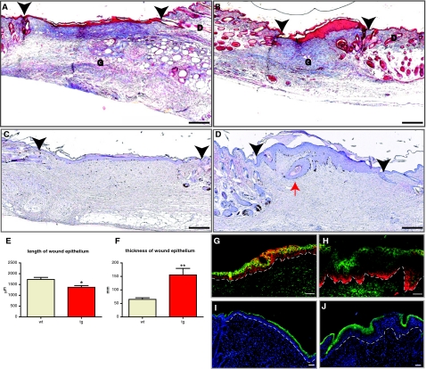
Reduced size, hyperthickened epithelium, and normal epidermal differentiation in 14-day excisional wounds. Six wild-type and six transgenic animals (nine wounds for each genotype) were analyzed. A and B: Masson trichrome-stained sections from the middle of representative 14-day excisional wounds of wild-type (A) and transgenic (B) animals are shown. Granulation tissue (G) and dermis (D) are indicated. C and D: BrdU-stained sections from the middle of representative 14-day wounds of wild-type (C) and transgenic (D) animals are shown. Black arrowheads indicate the edges of the original wound. The red arrow points to an epidermal cyst. E: Graphic representation of the average length of wound epithelium (*P = 0.0099). F: Graphic representation of the average thickness of wound epithelium (**P = 0.0014). G and H: Immunofluorescence staining of the 14-day wound epithelium in wild-type (G) and transgenic (H) mice, using a keratin 14 antibody (red) and a keratin 10 antibody (green). I and J: Immunofluorescence staining of 14-day wound epithelium from wild-type (I) and transgenic (J) mice, stained with a loricrin antibody (green) and DAPI (blue). Dotted line identifies the location of a basement membrane. Scale bars: 200 μm (A–D); 50 μm (G–J). Original magnifications: ×10 (A–D); ×20 (G–J).

