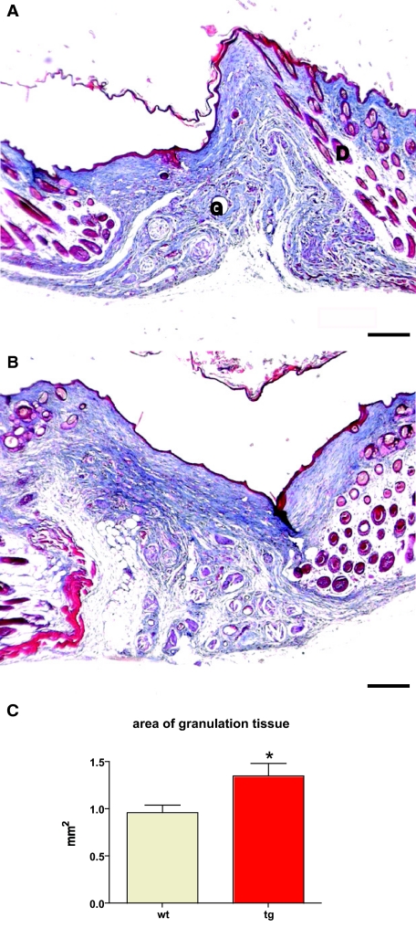
Figure 6.
Normal epithelial thickness and general granulation tissue architecture in 21-day excisional wounds. A and B: Masson trichrome staining of representative sections from the middle of 21-day excisional wounds taken from wild-type (A) and transgenic (B) mice. Granulation tissue (G) and dermis (D) are indicated. Five wild-type animals (10 wounds) and five transgenic animals (9 wounds) were analyzed. C: The area of granulation tissue (*P = 0.0205) is shown. Scale bars = 200 μm. Original magnifications, ×10.

