Figure 8.
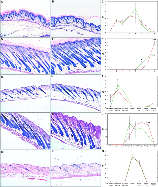
mIGF-1 accelerates late hair follicle morphogenesis and anagen development. Giemsa staining of back skin sections from wild-type (A, D, G, J, M) and transgenic (B, E, H, K, N) mice is shown. C, F, I, L, and O: Quantitative analysis of hair follicles from wild-type (green) and transgenic (red) mice in various stages of morphogenesis and cycling. *P ≥ 0.05 and ***P ≥ 0.001. A–C: One dpp, early morphogenesis. D–F: Eight dpp, late morphogenesis. G–I: Seventeen dpp, early catagen. J–L: Twenty-eight dpp, anagen. M–O: Forty-nine dpp, telogen. X-axes in C and F identify nine distinct hair follicle morphogenesis stages. X-axes in I, L, and O identify distinct stages of hair follicle cycling. Y-axes show the percentage of hair follicles in each distinct stage of hair follicle morphogenesis or cycling. Original magnifications, ×10.

