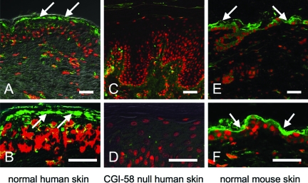
Figure 3.
CGI-58 Distribution in the Skin. A, B: Immunoreactivity of the anti-CGI-58 antiserum in normal human skin. CGI-58 immunolabeling (green, FITC) was seen in the epidermis, predominantly in the granular layer (arrows) and in the dermal fibroblasts in human skin. C, D: Anti-CGI-58 antiserum showed no labeling in CGI-58 null epidermis of a DCS patient with homozygous CGI-58 truncation mutation (Akiyama et al,4). E, F: CGI-58 distribution in murine skin. CGI-58 labeling (Green, FITC) was also observed in the epidermis and fibroblasts of mouse skin. CGI-58, green (FITC); nuclear stain, red (propidium iodide). Scale bars = 50 μm.

