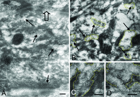
Figure 4.
Cryosubstituted, Cryofixed Postembedding Immunoelectron Microscopy with Anti-CGI-58 Antiserum in Normal Human Epidermis. A: Anti-CGI-58 antibody deposition labeled with 5 nm gold particles was seen on cytoplasmic vesicles (arrows) in the uppermost spinous and granular layer keratinocytes. CGI-58 was secreted with lamellar content of a vesicle to the intercellular space (open arrow). B: CGI-58-positive granules (arrows) contained lamellar structures inside and were identified as LGs. C, D: CGI-58 labeling was observed on the lamellar structure of LGs. Margin of LGs is traced with yellow dots. Scale bars = 100 nm (A–D).

