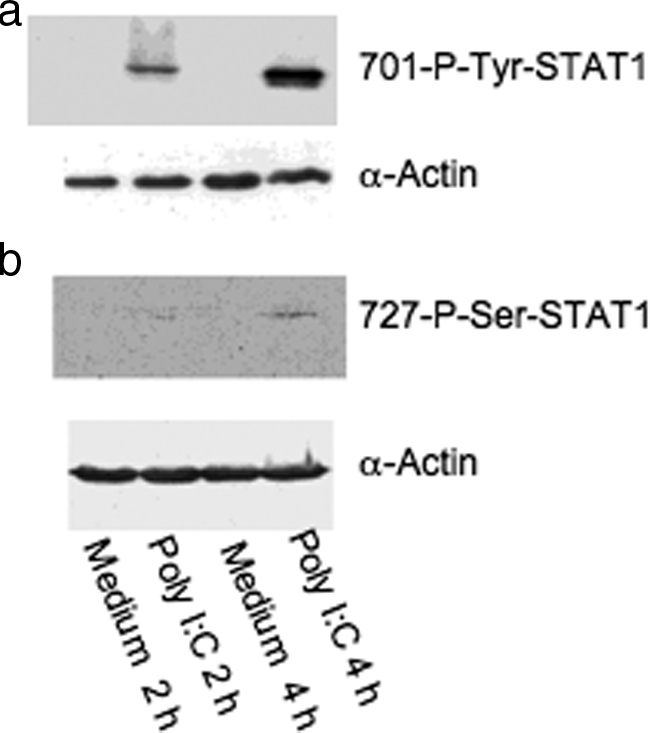
Figure 2.

Phosphorylation of STAT1 by Western blots. M-SMCs were serum-starved for 18 hours and then treated for 2 or 4 hours with poly I:C in 2% serum-containing medium as indicated and equal protein (30 μg) amount of cellular extracts were used for Western blot analyses using phosphor-specific antibodies 701-tyrosine (a) and 727-serine (b) residues of STAT1 protein. The same blot was then reused with α-actin antibody to compare equal loading of each sample.
