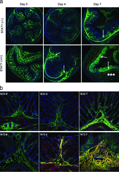
Figure 5.
Reduced HA deposition and less inflammatory changes in STAT1−/− mice after DSS treatment. a: Confocal image demonstrates HA polymers (green) in mouse colon sections after DSS treatment in STAT1−/−-null (top) and STAT1+/+ wild-type animals (bottom). The null mice section demonstrates minimal staining on day 4 and moderate staining on day 7 that is localized to the submucosa (arrow), whereas the wild-type mice have moderate staining of the submucosa (arrow) on day 4 and heavy staining of the lamina propria (asterisks) on day 7; cells are identified by their nuclei (blue). b: HA (green) and IαI (red) staining in colon designated as wild-type STAT1 (WT) and STAT1-null mice (KO) sections after a 4- or 7-day treatment with DSS.

