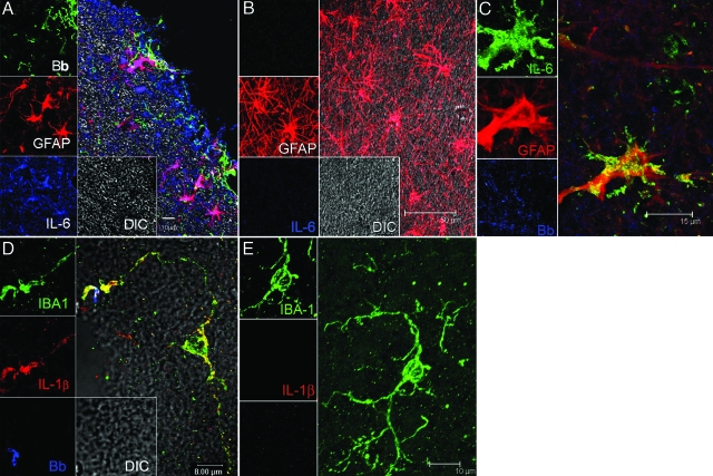
Figure 1.
Visualization of the production of cytokines IL-6 and IL-1β by glial cells in B. burgdorferi-exposed frontal cortex tissue explants and brain sections from animals given stereotaxic inoculations with B. burgdorferi. A: IL-6-producing astrocytes appear pink because of co-localization of IL-6 antibody (labeled blue) with antibody to the astrocyte marker GFAP (red). The spirochetes stained with fluorescein isothiocyanate-labeled B. burgdorferi antibody (Bb) appear green. Unstained tissue appears gray under differential interference contrast (DIC). B: Astrocytes labeled with GFAP (red) in brain tissue slices incubated in medium plus brefeldin A in the absence of spirochetes had no detectable IL-6. C: Visualization of IL-6-producing astrocytes in a tissue section taken at the site of inoculation from an animal given stereotaxic inoculations with live B. burgdorferi. IL-6-producing astrocytes appear yellow because of co-localization of antibody to IL-6 (green) and antibody to the astrocyte marker GFAP (red) in the vicinity of B. burgdorferi antigen stained blue. D: IL-1β-producing microglia appear yellow because of co-localization of antibody to the microglial marker IBA 1 (green) and antibody to IL-1β (red). Spirochetes in this image appear blue. E: Microglia labeled with IBA-1 (green) in brain slices incubated in medium plus brefeldin A in the absence of spirochetes had no detectable IL-1β.

