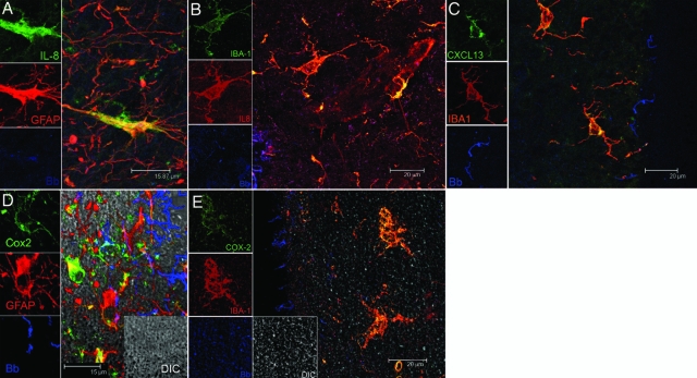
Figure 2.
Visualization of the production of IL-8, CXCL13, and COX-2 by glial cells in B. burgdorferi-exposed frontal cortex tissue explants. A: IL-8-producing astrocytes appear yellow because of co-localization of IL-8 antibody (green) and antibody to astrocyte marker GFAP (red). B: IL-8-producing microglia appear yellow because of co-localization of IL-8 antibody (red) and antibody to microglial marker IBA 1 (green). C: Production of CXCL13 in microglia appears yellow because of overlap of antibody to CXCL13 (green) and IBA 1(red). D: COX-2-producing astrocytes are seen as yellow because of overlap of GFAP-staining astrocytes (red) and COX-2 (green). E: COX-2-producing microglia are seen as yellow because of overlap of IBA-1-staining microglia (red) and COX-2 (green). Spirochetes appear blue in all sections.

