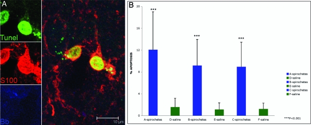
Figure 4.
Oligodendrocyte apoptosis observed in rhesus CM60 given stereotaxic inoculations with live B. burgdorferi. A: Apoptotic oligodendrocytes show TUNEL-positive nuclei stained green. Oligodendrocytes appear red because of staining with antibody to S-100. B. burgdorferi antigen appears blue in color. B: Percentage of oligodendrocytes (S-100-positive, GFAP-negative) undergoing apoptosis in brain sections from the sites of spirochetal inoculation (A–C) and corresponding control sites (D–F) as quantified by the in situ TUNEL assay.

