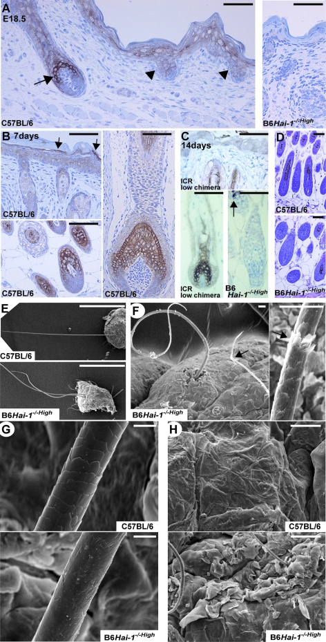
Figure 6.
Abnormalities of hair and whiskers in Hai-1/Spint1-deleted mice. A: Expression and localization of Hai-1/Spint1 protein in wild-type skin (C57BL/6) at E18.5. Hai-1/Spint1 is strongly expressed by hair cortex cells of early anagen hair follicle (arrow). It is also expressed in epidermal keratinocytes, especially in the granular layer, and to a lesser degree by primary hair germ (arrowheads). The skin of Hai-1/Spint1-deleted mouse (B6Hai-1−/−High, E18.5) was simultaneously immunostained as a control (right). B: Expression of Hai-1/Spint1 in wild-type skin at day 7. Hai-1/Spint1 is strongly expressed by hair cortex and cuticle cells and weakly by inner root sheath of follicles (bottom left, dorsal skin pelage hair follicles; right, whisker follicle). In the epidermis, immunoreactivity for Hai-1/Spint1 is also observed and the expression appears to be most evident in transitional cells between granular and corny layers (arrows). C: Expression of Hai-1/Spint1 in wild-type skin (ICR, chimerism 0%) at day 14. Hai-1/Spint1 is strongly expressed by hair cortex cells of anagen hair follicle. No immunoreactivity was detectable in B6Hai-1−/−High hair follicles. Arrow indicated melanin pigments of black hair. D: Histology (H&E) of anagen hair follicles of dorsal skin pelage hair at 1 week after birth. E–H: Scanning electron microscopy images of whiskers and hair (E-–G) and skin surface (H) of wild-type (C57BL/6) and B6Hai-1−/−High mice. Fragile appearance (F, arrows) and poorly formed cuticle layer (G) are observed in B6Hai-1−/−High mouse. Scale bars: 1 mm (E); 20 μm (F); 10 μm (G); 50 μm (A–D, H).

