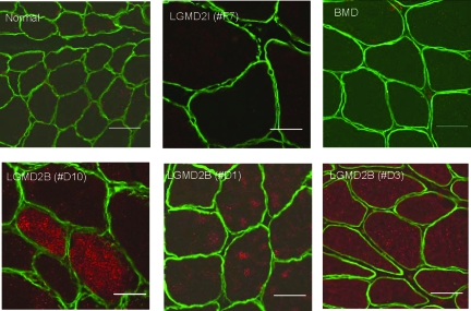
Figure 2.
Rab27A Immunostaining in Muscle Biopsies. Shown are confocal images of Rab27A (red) and merosin (laminin α2) (green) co-stained in patient muscle biopsies. Dysferlin-deficient patient muscle (LGMD2B) showed high-level but variable expression of Rab27A in myofibers. In LGMD2B Patient #D10, a subset of myofibers showed high level Rab27A expression. In Patients #D1 and #D3, Rab27A was seen at lower levels in the majority of fibers. Rab27A signal was not detectable in normal controls, LGMD2I, or Becker muscular dystrophy. Scale bar = 50 μm.

