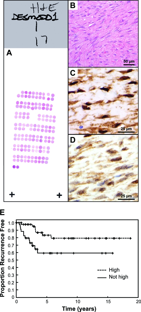
Figure 3.
A: Representative H&E-stained slide from one of the three TMA blocks. B: H&E of desmoid tumor on the TMA. Immunohistochemistry shows high (C) and moderate (D) levels of nuclear β-catenin. Nuclear reactivity was considered high (=3) when the immunoreactivity completely precluded visualization of the nuclear hematoxylin counterstain whereas moderate (=2) and weak (=1) staining allowed such visualization to varying degrees. This determination was made at ×200 magnification with advancement to ×400 when necessary for cases with minimal reactivity. E: Kaplan-Meier analysis of time to recurrence from primary surgery for desmoid tumor in patients (n = 84) in which the actual primary tumors were available for TMA immunohistochemistry. Desmoid tumors showing attenuated nuclear β-catenin intensity levels [moderate (=2), minimal (=1), or rarely, negative (=0)] demonstrated by immunohistochemistry increased propensity to recurrence when compared to desmoids with high (=3) levels (P = 0.0406). Moderate, minimal, and negative staining groups were combined (not high) because they behaved similarly and because there were few patients in the minimal and negative groups. Original magnifications: ×200 (B); ×400 (C, D).

