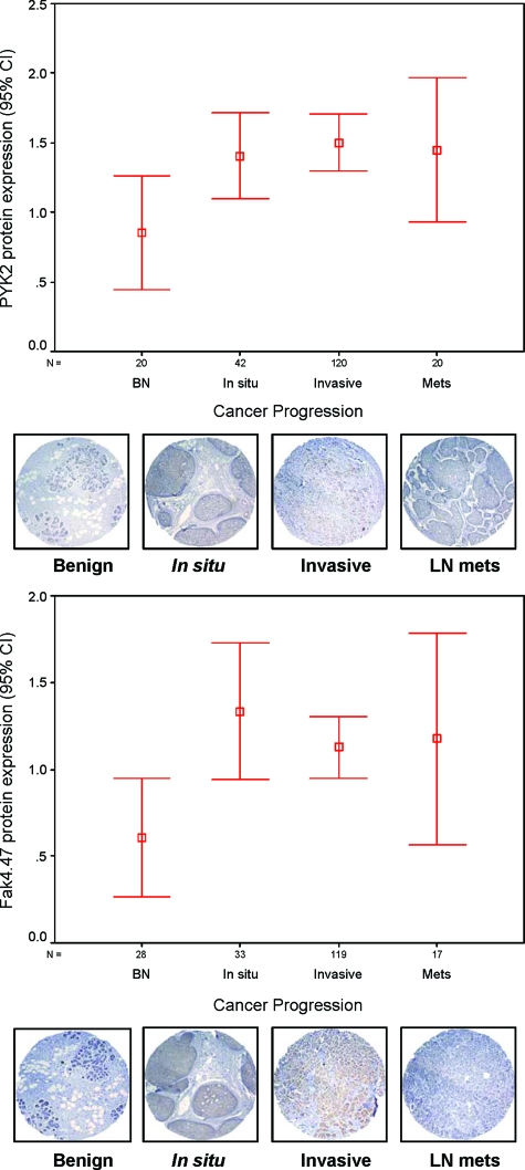
Figure 1.
Summary of Pyk2 and FAK expression in progressive breast cancer tissue array specimens. Pyk2 and FAK protein expression in a TMA of benign breast tissues versus carcinoma in situ, invasive, and lymph node metastasis, expressed in percentages. Confidence intervals (95%) show normalized mean protein intensity units of Pyk2 and FAK as determined by quantitative evaluation of immunohistochemistry; scale bars, ±SE. BN, benign; In situ, in situ breast carcinoma; Invasive, invasive breast cancer; Mets, lymph node breast metastasis. Error bars are representative cores of immunostaining of Pyk2 and FAK expression in various stages of breast cancer progression using TMA. Benign breast epithelium showed negative or very low intensity compared with different stages of breast cancer. *P < 0.05 in situ, invasive versus benign.

