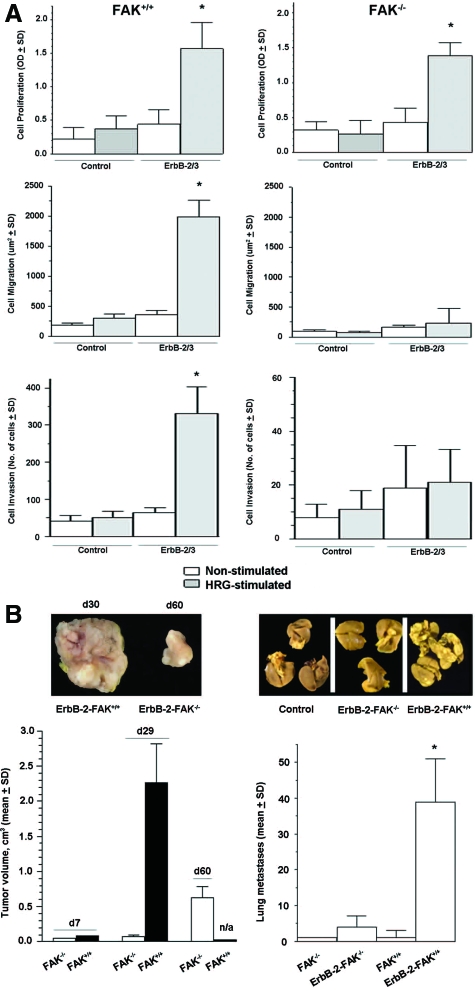
Figure 3.
ErbB-2/3 receptor overexpression partially rescues tumorigenesis defects of FAK−/− cells. A: Cell proliferation was measured using 3-(4,5-dimethylthiazo-2-yl)-2,5-diphenyltetrazolium bromide metabolic assay on exponentially growing cells after 48 hours of HRG stimulation and nonstimulated controls. Cell migration was determined by the phagokinetic track assay, where cells were allowed to migrate on gold colloidal particle-covered coverslips in the presence or absence of HRG. Total cell motility was quantified by processing the average area free of gold colloidal particles in square millimeters ± SD. Cell invasion was evaluated by culturing cells on Boyden chambers coated with matrigel and stimulating them for 48 hours using HRG as chemotactic agent in the lower chamber as described in Materials and Methods. The number of invading and hematoxylin stained cells were counted, and the percentage of invading cells was determined. Values are means ± SD from three independent experiments (P < 0.05, ErbB-2/3 versus control). *P < 0.01, ErbB-2/3 FAK−/− versus control FAK−/−; P < 0.001, ErbB-2/3 FAK+/+ versus control FAK+/+ B: Exponentially growing cells (1.0 × 10⁶) were injected subcutaneously into the flank of SCID mice (left). Tumor growth was monitored over time as indicated in Materials and Methods. Each point represents the average of 8 animals ± SEM. Tumor volumes are not applicable for FAK+/+ at day 60 because animals were sacrificed on day 29 because of debilitating large tumors. Exponentially growing cells (1.0 × 10⁶) were inoculated intravenously to SCID mice (right). Mice were sacrificed on day 42 after inoculation, and lungs were fixed in 10% Bouin’s fixative. Lung surface metastases were counted using a stereomicroscope. Each bar represents the average lung metastases (n = 8) ± SEM. *P < 0.005, ErbB-2/3 FAK−/− versus ErbB-2/3 FAK+/+.

