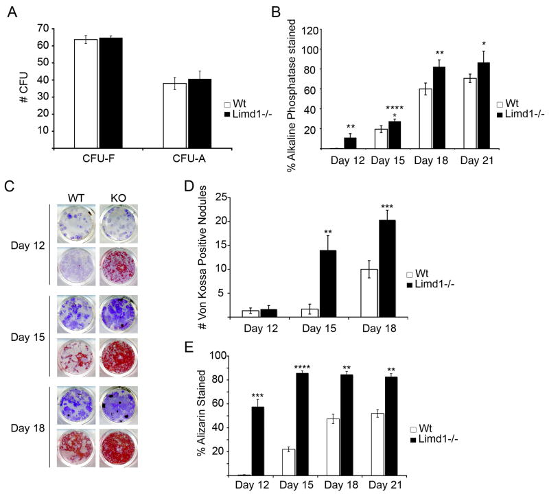
Fig. 4.
Limd1−/− mice have increased numbers of osteoblast progenitors.
(A) A comparison of the number of hematoxylin positive fibroblast colony forming units (CFU-F) or Oil Red O positive adipocyte colony forming unit (CFU-A) present after culturing Limd1−/− and Wt mice bone marrow stromal cells in normal or adipogenic media for 9 days. (B) A comparison of the number of alkaline phosphatase positive CFU-O present after culturing Limd1−/− and wt bone marrow stromal cells in osteogenic media for the number of days indicated. (C–E) Limd1−/− and Wt marrow stromal cells were cultured in osteogenic media for the number of days indicated days then fixed and stained for the presence of alkaline phosphatase (purple, all wells). Mineralization was visualized by Von Kossa (black, top row for each time point) or Alizarin Red S (red, bottom row for each time point) and the number of Von Kossa positive CFU-O (D) or Alizarin positive CFU-O (E) enumerated. *Indicates a significant difference between Limd1−/− and Wt (*p<0.05, **p<0.01, ***p<0.005, ****p<0.001, as determined by the student’s paired t-test).

