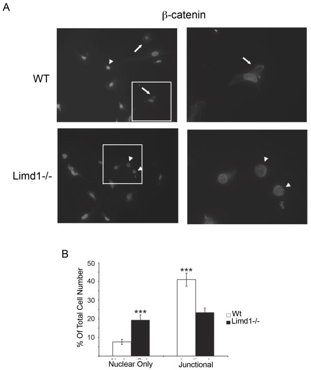
Fig. 5.
Limd1−/− calvarial cells have more nuclear β-catenin than Wt controls.
(A) Primary Wt or Limd1−/− calvarial osteoblasts were fixed, permeabilized and stained for β-catenin and DAPI. Cells with nuclear only β-catenin (arrowheads) and junctional β-catenin (arrows) are highlighted. (B) The percentage of cells with nuclear only β-catenin and the percentage of cells containing junctional β-catenin was determined and graphed. (***p<0.005, as determined by the student’s paired t-test).

