
Figure 4.
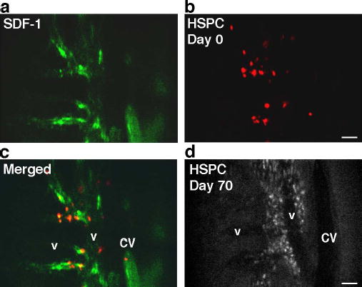
HSPC homing to SDF-1-positive vascular microdomains. a–c. Co-localization of SDF-1 (green) and HSPC (red) 2h post-injection (parasagittal region of area 3). d. The same mouse imaged 70 days post-HSPC injection (approximately the same area as in a–c). Although fluorescence signal intensity is diminished due to HSPC proliferation, engrafted cells are apparent in the perivascular spaces. Scale bars = 100 μm. CV=central vein, v=venule.