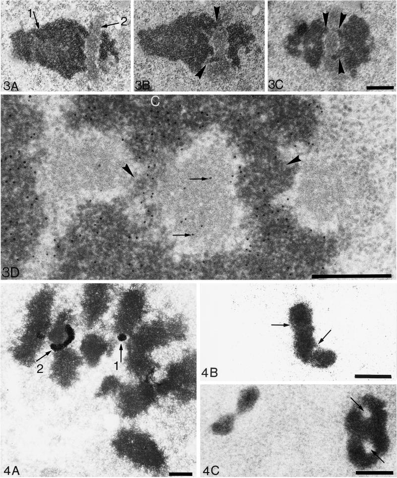
Figure 3.
Ultrastructure of crescent-shaped NORs. Crescent-shaped NORs observed in ultrathin sections of acetylated Ehrlich cancerous cells blocked in metaphase. (A–C) Three serial tangential sections parallel to the long axis of one NOR-bearing chromosome. After uranyl and lead counterstaining, chromatin (arrow 1) appears with a high contrast to electrons, whereas the fibrillar component of one NOR shows a lower contrast (arrow 2). On A, the elongated NOR (240 nm in diameter) is interrupting the chromosome. However, on two serial sections of this structure (B and C), two fibers of chromatin 100–150 nm in diameter (arrowheads) appear in continuity with the left and right sides of the chromosome. This suggests that one elongated NOR is crossed by two bridges of chromatin (one bridge per chromatid) and does not interrupt the continuity of the chromosome. Consequently, on some well-oriented sections, the NOR seems to be divided in three distinct parts as observed on C. Bar, 500 nm. (D) Specific detection of DNA by the TdT method on an ultrathin section of an acetylated Ehrlich cell blocked in metaphase. The elongated NOR shown is similar (but is turned at 90°) to the one in A–C. Thus the three fibrillar zones belong to the same NOR (1800 nm long and 400 nm thick) that is divided in three parts by two bridges of chromatin (arrowheads) as above (C). Gold particles, 10 nm in diameter, specifically reveal DNA. Condensed chromatin (labeled C) is labeled with the highest density of gold particles. Moreover, a significant number of gold particles is localized on the NOR attesting for the presence of uncondensed DNA (arrows). Note that there is no background. Bar, 500 nm.

