
Figure 1.
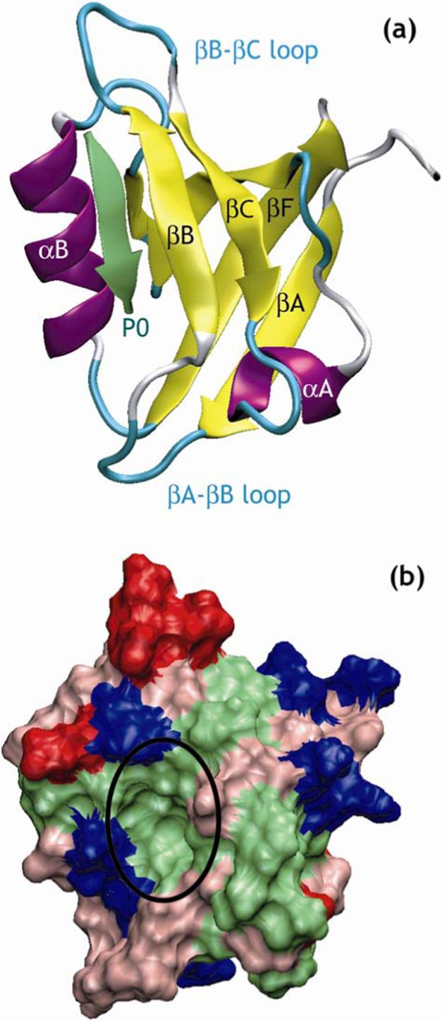
Representative structure of a PDZ domain in complex with its target peptide. (a) Ribbon representation of the crystal structure of PDZ3 of PSD-95 in complex with a peptide in its binding site 3.
The peptide is represented in green and the C-terminus of the peptide is indicated by P0 (b) Molecular surface representation of PDZ3 of PSD-95 with basic residues in blue, acidic residues in red, polar residues in pink and hydrophobic residues in green. The binding pocket for the peptide is indicated by a black circle. Figures created using VMD 92.