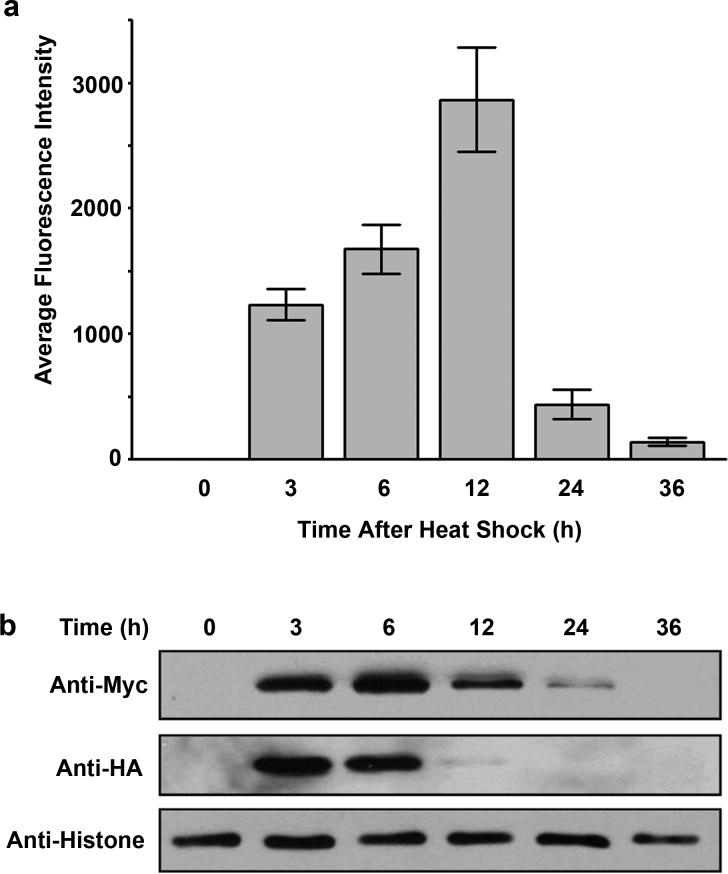
Figure 3. Fluorescence and expression profiles of the bFOS-bJUN interaction.
(a). Ten synchronized young adult worms carrying the plasmids pCE-bFOS-VC155 and pCE-bJUN-VN173 were heat-shocked at 33°C for 3 h, followed by incubation at 20°C. At the indicated time points after the heat shock, fluorescent images were acquired using the YFP filter setup (20X objective lens). The fluorescence intensity in the nucleus of five intestinal cells from each worm was quantified, and the average fluorescence intensity from 10 worms was calculated and presented as a function of time. Error bars indicate standard deviation. (b). Immunoblotting analysis of expressed HA-bFOS-VC155 and Myc-bJUN-VN173 fusion proteins at the indicated time after heat shock.

