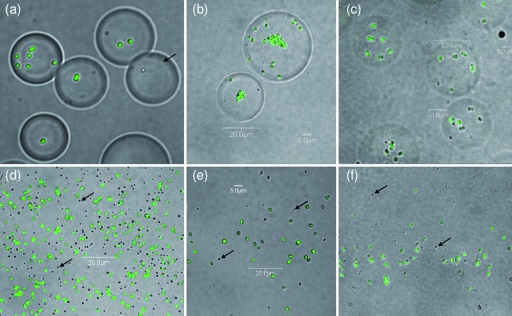
FIG. 2.
Representative confocal microscopic images of particles collected in the Ultimate cascade impactor from an aerosol (12-μm MMAD) generated by the FFAG containing FluoSpheres (a and d), B. atrophaeus spores (b and e), or Escherichia coli MRE162 cells (c and f). The imaged particles are 8 to 16 μm (a, b, and c) or 1 to 2 μm (d, e, and f). Particulates were aerosolized at a concentration of 109 ml−1. Arrows indicate empty particles.

