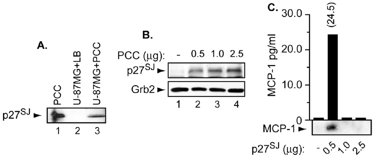
Figure 1. Expression of p27SJ under normal physiological conditions and the status of MCP-1.
A, B. U-87MG (A) and microglial (B) cells were treated with purified callus culture of H. perforatum. Fifty micrograms of cell lysates were subjected for Western blot analysis using anti-p27SJ rabbit polyclonal antibody. Arrows depict the position of p27SJ protein. Anti-Grb2 was used for equal protein loading. C. MCP-1 levels were measured in supernatants collected from microglial cells treated with an increasing amount of p27SJ by ELISA and or Western blot. Number on the top of the bar represents the amount of MCP-1 secreted in the presence of p27SJ as measured by ELISA. Arrow depicts the position of MCP-1 protein. Histogram represents amount of MCP-1 secreted following p27SJ treatment.

