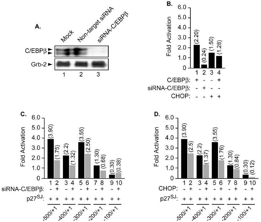
Figure 4. Functional interplay between C/EBPβ and p27SJ in the presence of CHOP and siRNA-C/EBPβ.
A. Microglial cells were transfected with non-target siRNA or with siRNA-C/EBPβ as described in Materials and Methods. Cellular proteins were then prepared and subjected to Western blot analysis using anti-C/EBPβ antibody. Arrows point to the positions of C/EBPβ proteins. Equal loading of proteins was controlled using anti Grb-2 antibody (lower panel). Next, microglial cells were transfected with 0.5 µg of the MCP-1 promoter alone or in the presence of CHOP (B, D) or siRNA-C/EBPβ (B, C). Four hours post transfection, the cells were infected with adeno-p27SJ at an MOI of 1 (C, D) or adeno-null (data not shown). CAT activity was determined 24 h after transfection. The values shown on the top of each bar represent the fold activation over the basal promoter activity which is arbitrarily set at one. The data represent the mean value of at least three separate transfection experiments.

