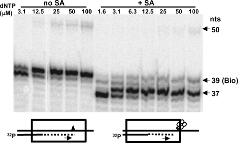
Figure 5. Stalling of primer extension by HIV-1 RT due to a biotin residue placed at a specific position in the template in the absence or presence of SA.
(Left panel) Five nM 5′-[32P]-L20 primer annealed to WL50-Bio39 template was extended with 200 nM RT and various concentrations of dNTP (indicated above the lanes) and 6 µM biotin for 45 min at 37°C. (Right panel) 5′-[32P]-L20/WL50-Bio39 P/T was first incubated for 2-3 min with 100 nM SA at 37°C and then RT, dNTPs and biotin were added and primer extension was carried out as above. Labeled products were fractionated on 20% polyacrylamide under denaturing conditions. Arrows indicate 50 nt (full length of template), 39 nt (primer extended to the position of the biotin residue), and 37 nt (primary stop site for primer extension in the presence of SA.). Diagrams at the bottom of the figure show the direction of primer extension (arrows) and the likely position of RT (box) after stalling on a template containing the biotin residue (triangle) in the absence (left) or presence (right) of SA (shown as a tetramer of circles).

