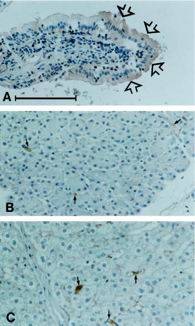
Figure 6.
Distribution of galectin-4 in a pig small intestinal villus (A), pancreas (B), and liver (C). Note that the enterocytes, in particular those at the tip of the villus (arrows in A) are clearly labeled. In the two other tissues the epithelial paranchyma is unstained although some immunoperoxidase staining may be seen in relation to stromal elements (small arrows). Bar, 100 μm.

