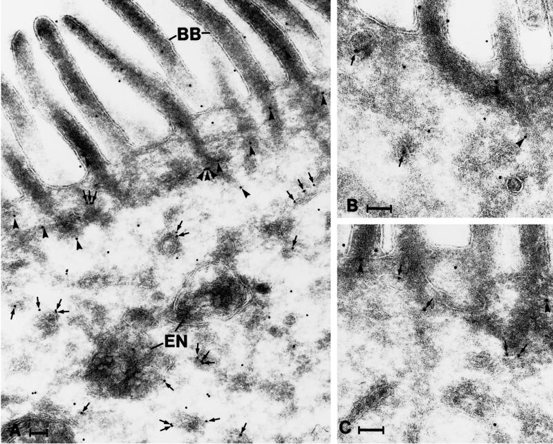
Figure 7.
Distribution of galectin-4 in the apical portion of enterocytes as revealed by immunogold labeling. Gold particles are seen throughout the cytoplasm, in association with tubulo-vesicular structures (arrows) and the actin rootlets of microvilli (arrowheads). Note that many of the gold particles associated with tubulo-vesicular structures are found on their cytosolic surface. BB, brush border; En, endosomes. Bar, 100 nm.

