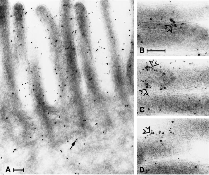
Figure 8.
Clustering of galectin-4 and aminopeptidase N. Immunogold double labeling for aminopeptidase N (5-nm gold) and galectin-4 (10-nm gold). (A) Aminopeptidase N and galectin-4 are present in the brush border, but little aminopeptidase N is seen in the cytoplasm (arrow). (B–D) Galectin-4 and aminopeptidase N often form clusters in the microvillar membrane (open arrows). Bars, 100 nm.

