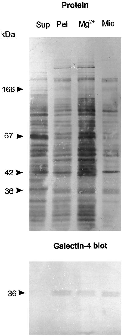
Figure 9.
Subcellular distribution of galectin-4. About 1 g of frozen intestinal mucosa was thawed and homogenized in a Potter-Elvehjem homogenizer in 10 ml of 25 mM HEPES and 150 mM NaCl, pH 7.0. The homogenate was cleared by centrifugation at 500 × g for 5 min and then centrifuged at 48,000 × g for 30 min, to obtain a pellet (Pel) and a supernatant (Sup). The pellet was resuspended in 10 ml of the above buffer and 50 μl of this fraction and of the supernatant was analyzed by SDS-PAGE. Likewise, from 1 g of intestinal mucosa, Mg2+-precipitated (Mg2+) and microvillar (Mic) fractions were prepared and resuspended in equal volumes of 25 mM HEPES and 150 mM NaCl, pH 7.0, and samples of 50 μl were examined by SDS-PAGE. After electrophoresis, the gel tracks were Western blotted with the galectin-4 antibody and afterward stained for protein with Coomassie brilliant blue.

