Figure 1.
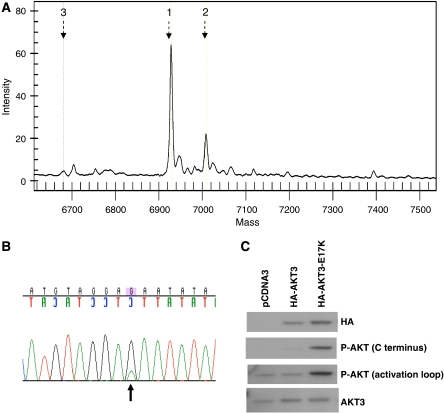
AKT3 E17K mutation in melanoma. (A) Mass spectroscopy-based detection of AKT3 E17K mutation in a human melanoma clinical specimen. Peaks correlating with wild-type AKT3 (‘1’) and mutant AKT3 (‘2’) are indicated. (‘3’=predicted mass of unincorporated primer). Only the wild-type peak was seen in normal tissue from the same patient (Supplementary Figure 1). (B) Confirmatory Sanger sequencing of tumour analysed by mass spectroscopy-based method in (A). The missense substitution resulting in the E17K mutation is indicated with an arrow. (C) Western blotting analysis of A375 human melanoma cells transfected with empty control vector (‘pcDNA3’), HA-tagged wild-type AKT3 (‘HA-AKT3’), and HA-tagged mutant AKT3 (‘HA-AKT3 E17K’). Results shown are for cells growing under normal tissue culture conditions.

