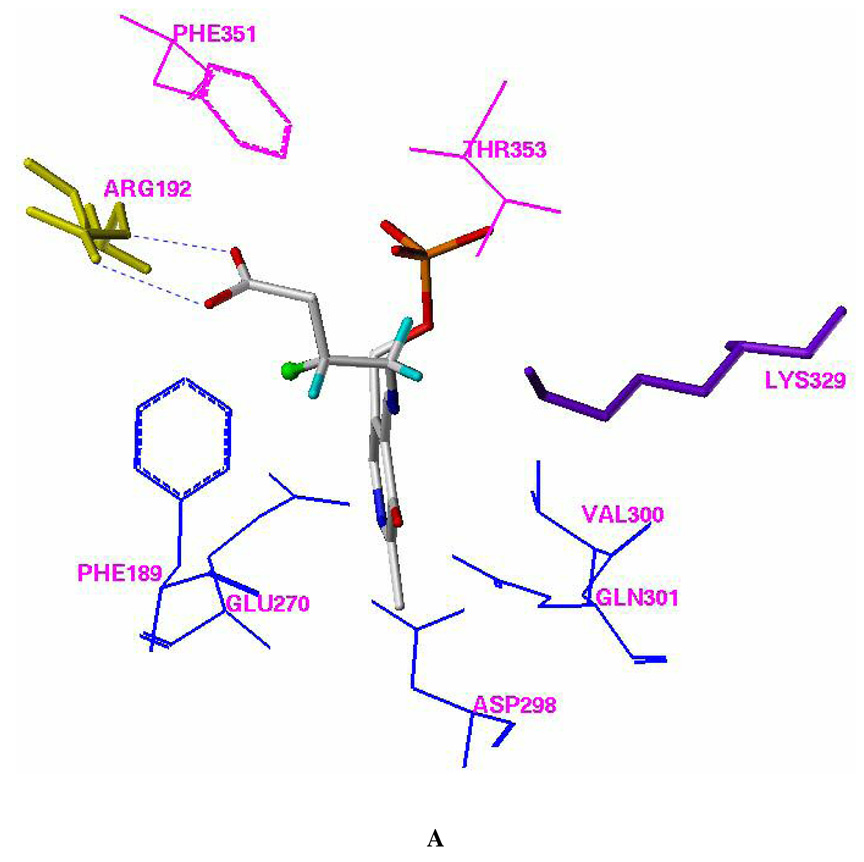
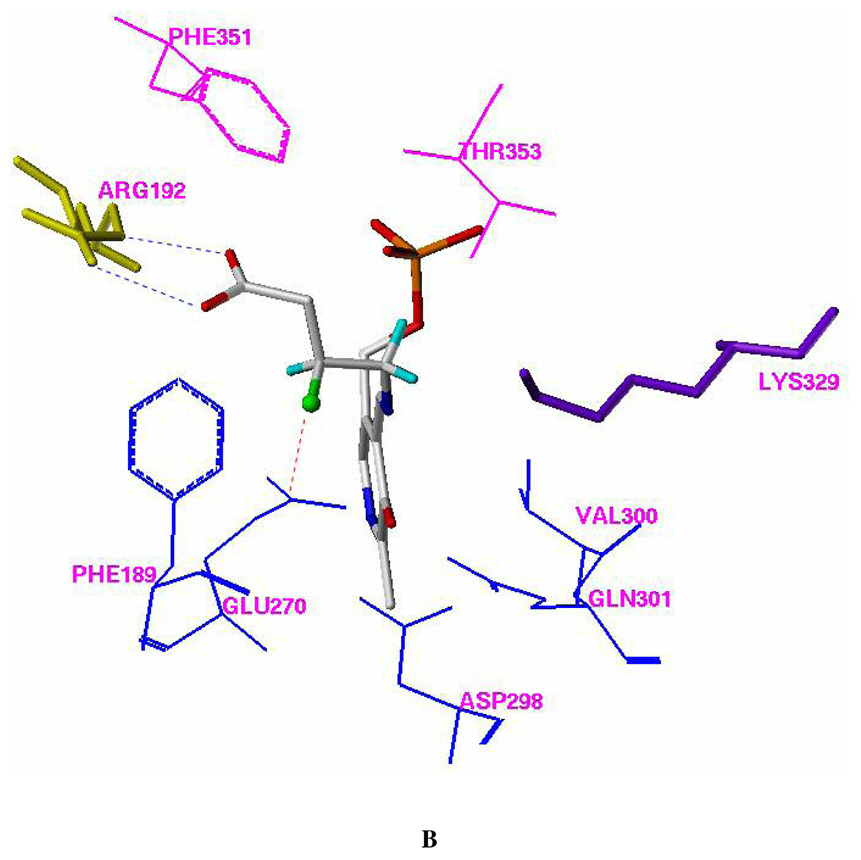
Figure 1.


Docking models of (R)-2 (A) and (S)-2 (B) with pig liver GABA-AT (FlexX model). Residues Phe351 and Thr353, which are from the other monomer of the homodimer, are shown in magenta. All of the hydrogen atoms except those attached to C-3 and C-4 of (R)-2 and (S)-2 were omitted for clarity. The fluorine atom is presented in a ball and stick model. The putative H-bonds between Arg 492 and the carboxylic group of (R)-2 or (S)-2 are shown in blue. The putative dipole-dipole interaction (C δ+-Fδ−…Cδ+-O−) between the side chain of Glu270 and the C-F group of (S)-2 is shown in red.
