Figure 2.
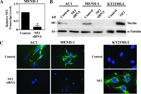
In vitro model system of merlin expression in human meningeal cells. Endogenous merlin was silenced in AC1 and MENII-1 cells using NF2 specific siRNA. In parallel, merlin isoform 1 was exogenously expressed in KT21MG1 cells using retroviral mediated gene transfer. (A) NF2 transcript levels were measured by quantitative PCR and showed a 5.6-fold reduction in MENII-1-NF2-siRNA cells compared with MENII-1-Control cells. Asterisk denotes statistical significance (P < .05). (B) Western blot analysis of cell lysates derived from AC1, MENII-1, and KT21MG1 stable cell populations was used to confirm loss or gain of merlin. Whereas merlin expression was observed in AC1-Control and MENII-1-Control cells, NF2 siRNA abolished expression of merlin in AC1-NF2-siRNA and MENII-1-NF2-siRNA cells. In parallel, KT21MG1-Control cells lacked merlin, whereas KT21MG1-NF2 cells expressed wild type merlin. Levels of α-tubulin were determined in the same samples as a loading control. Immunoblot of one representative experiment of three with similar results is shown. (C) Immunofluorescence using the A19 polyclonal antibody against merlin revealed the presence of cytoplasmic staining in AC1-Control and MENII-1-Control cells and its absence in AC1-NF2-siRNA and MENII-1-NF2-siRNA cells. In contrast, KT21MG1-Control cells had no staining, whereas KT21MG1-NF2 cells had cytoplasmic staining. Merlin immunolabeling is shown in green, and nuclear DAPI counterstaining is shown in blue.

