Figure 5.
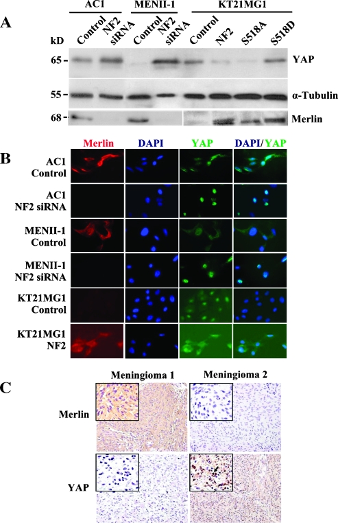
Protein levels of YAP are up-regulated and localized to the nucleus in NF2-deficient cells. (A) Total cell lysates were subjected to Western blot using a YAP- or merlin-specific antibody. Increased YAP protein expression was observed when NF2 was suppressed in AC1 and MENII-1 cells compared with controls. Conversely, exogenous expression of merlin decreased YAP in KT21MG1 cells compared with controls. Expression of a nonphosphorylated, active merlin (S518A NF2) was also associated with lower levels of YAP compared with the expression of pseudophosphorylated inactive merlin (S518D NF2). Levels of α-tubulin were determined in the same samples as loading control. Results were reproduced in three independent experiments. (B) YAP was translocated to the nucleus in merlin-deficient cells. Immunofluorescence staining was used to show that YAP was localized to the nucleus in AC1-NF2-siRNA, MENII-1-NF2-siRNA, and KT21MG1-Control cells. In contrast, YAP was primarily cytoplasmic in AC1-Control, MENII-1-Control, and KT21MG1-NF2 cells. Merlin immunolabeling is shown in red; YAP staining is shown in green; and nuclear DAPI counterstaining is shown in blue. (C) In situ immunostaining of merlin and YAP in serial sections of primary human meningioma tumors. We surveyed 37 primary meningiomas by immunohistochemistry. YAP expression was minimal to absent in 95% of merlin-positive meningiomas (a representative tumor is shown here as Meningioma 1). In contrast, YAP was expressed and localized to the nucleus in 92% of merlin-negative meningiomas (a representative tumor is shown here as Meningioma 2). Arrow depicts an example of YAP nuclear localization. Insets show images at higher magnification.

