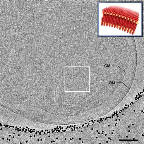
Fig. 1.
Cryo-electron microscopy of Tsr chemoreceptor assemblies in whole E. coli cells. Shown is a projection image recorded from a plunge-frozen E. coli cell engineered to overproduce TsrQEQE (in the absence of serine) by using low-dose cryo-electron microscopy. The projection image shows patches of chemoreceptor assemblies in the native cytoplasmic membrane (CM) contained within a cell with an intact outer membrane (OM). The cell is close to the edge of a hole in a holey carbon grid containing vitreous ice; the black dots are 15-nm gold particles added for use as fiducial markers in tomogram reconstruction. (Inset) A schematic perspective view of “zipper”-like chemoreceptor assemblies from a region such as that enclosed by the white box. (Scale bar: 100 nm.)

