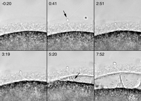
Figure 8. Heparin blocks sperm entry.
The fertilization process in the heparin-injected egg was monitored with a CCD camera, and the key moments were presented by still-shot photomicrographs. The moment of sperm attachment to the egg surface was set to t = 0:00 (min:sec). At 0:41, the sperm was still attached to the jelly coat (arrow). Afterwards, the sperm attempts but fails to penetrate the jelly coat. At 5:20, the vitelline layer is visibly elevated, but the sperm is still completely outside the jelly coat. The formation of the fertilization cone is evident under the elevating membrane (arrow). At 7:52, the vitelline layer is further elevated, but the fertilization cone fails to pull in the sperm head. The fertilization envelope is now being established while the sperm is still outside. The motion picture of the entire process is available as a video file in Data S3.

