
Figure 1.
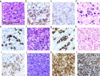
Histology of representative ALCL and pre-B-ALL cases. All panels shown at identical magnification. a–e: ALK+ ALCL stained with H&E (a), immunohistochemistry with anti-CD2 (b), anti-CD30 (c), in situ hybridization for EBV (d), or anti-ALK (e). f–i: ALK− ALCL stained with H&E (f), anti-CD3 (g), in situ hybridization for EBV (h), or anti-CD30 (i). j–l: pre-B-ALL stained with H&E (j), anti-CD79a (k), or anti-TdT (l). Images were captured with a Nikon Eclipse E1000 microscope (Nikon, Tokyo, Japan) using a 60×/0.95 objective lens (Nikon) and a Spot CCD camera (Diagnostic Instruments, McHenry, IL) using Spot software version 4.6 for image acquisition.