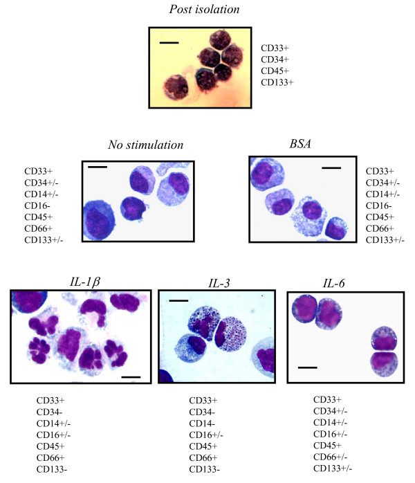
Figure 2.
Cytospin preparations of freshly isolated HPC and following culture for two weeks in non-stimulated, BSA or IL-stimulated EC supernatant. Freshly isolated HPC (Post isolation) with a dense nucleus and small cytoplasmatic rim increased up to two-fold in size and gained cytoplasma in non-stimulated and BSA-stimulated supernatants. With IL-1β stimulated supernatant they developed into hypersegmented cells and also into monocytic cells in part, with an increase in cytoplasma content. More than 50% of the cells stimulated with IL-3 developed eosinophilic granula, whereas cells in IL-6 stimulated supernatant resembled freshly isolated cells. Cells cultured in IL-6, BSA- and non-stimulated supernatants were still positive for CD34 and CD133. Diffquik staining, size bar 1 μm. magnifications ×200. One representative result of twelve independent experiments.

