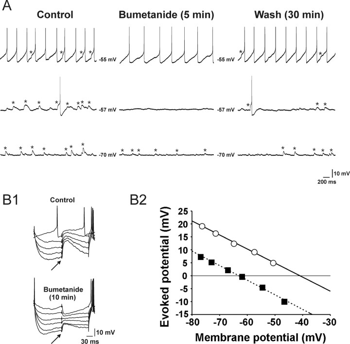
Figure 5.
Effects of bumetanide (10 μm) on EPSPs recorded in rat SCN neurons recorded with gramicidin-perforated-patch recording technique in the presence of dl-AP5 (100 μm) and DNQX (20 μm). A, Traces obtained at three different membrane potentials (i.e., −55, −57, and −70 mV) are presented to help demonstrate the bumetanide effect more clearly. Asterisks denote spontaneous EPSPs. B1, Synaptic potentials evoked at various membrane potentials by focal electrical stimulation (↑) of the dorsolateral border of the SCN in the absence (top) and presence of bumetanide (bottom). The membrane potential was varied by injecting a series of current pulses (duration, 250 ms; intensity, 0 to −0.06 nA). This cell exhibited spontaneous EPSPs (data not shown), as did the cell in A. B2, Estimation of the reversal potentials of the evoked synaptic responses shown in B1. The amplitudes of evoked responses are plotted against the baseline membrane potentials (○, control; ■, bumetanide). A linear regression was used to fit the data points. The intersections of the regression lines with the abscissa (−41 and −63 mV) were taken as the reversal potentials.

