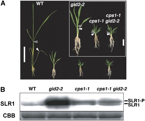
Figure 3.
Dwarfism and Accumulation of SLR1 Protein in gid2, cps1, and gid2 cps1.
(A) Gross morphology of the wild type, gid2-2, cps1-1, and the gid2-2 cps1-1 double mutant grown for 4 weeks. Closed and open arrowheads represent the uppermost positions of the 4th and 6th leaf sheath, respectively. Bar = 5 cm. Inset: close-up of the three mutants (bar = 1 cm).
(B) Top panel: protein gel blot showing the level of SLR1 protein accumulated in the seedlings shown in (A). SLR1-P, phosphorylated SLR1; SLR1, nonphosphorylated SLR1. Bottom panel, Coomassie blue (CBB) staining to show that approximately equal amounts of total protein (10 μg) were loaded in each lane.

