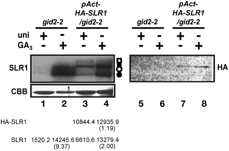
Figure 8.
No Change in the SLR1 Protein Level When SLR1 Is Expressed under the Control of a Constitutive Promoter in gid2-2.
HA-SLR1cDNA was expressed by the actin promoter under GA-rich or -deficient conditions in gid2-2. Left panel: SLR1 protein was detected by protein gel blot analysis with anti-SLR1 antibody. Open circle, phosphorylated SLR1; closed circle, nonphosphorylated SLR1; open square, phosphorylated HA-SLR1; closed square, nonphosphorylated HA-SLR1. Bottom panel: Coomassie blue (CBB) staining to show that approximately equal amounts of total protein (5 μg) were loaded in each lane. Right panel: HA-SLR1 protein was detected with anti-HA antibody. Total amount of protein in each lane was same as left panel. Numbers represent the band intensities (arbitrary units) of SLR1 and HA-SLR1 of each lane. Numbers in parentheses indicate the ratio of the band intensities between uni − GA3 + and uni + GA3 −.

