Figure 1.
PDV2 Is a Bitopic, Ring-Forming, Plastid-Targeted Protein with an IMS-Localized C Terminus.
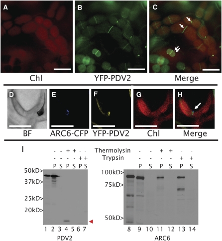
(A) to (C) Localization of YFP-PDV2 in epidermal cells of Arabidopsis young emerging leaves (∼4 to 6 mm long) expressing YFP-PDV2 under the control of the PDV2 promoter sequence. Single arrows point to midplastid PDV2 rings. Double arrows point to smaller plastids with peripheral and ring localization of PDV2.
(D) to (H) Colocalization of ARC6-CFP and YFP-PDV2 in the cells of young Arabidopsis leaves. Both proteins are expressed under the control of their native promoters.
(I) In vitro chloroplast import of [3H]Leu-labeled PDV2 or [35S]Met-labeled ARC6 and fractionation of chloroplasts following protease treatment and hypotonic lysis. In vitro–transcribed translation products are shown for PDV2 (lane 1) and ARC6 (lane 8). Pellet (P) fractions are shown in lanes 2, 4, 6, 9, 11, and 13. Supernatant (S) fractions are shown in lanes 3, 5, 7, 10, 12, and 14. The arrowhead points to the IMS-localized C-terminal fragment of PDV2 that remains following thermolysin treatment.
BF, bright-field image; ARC6-CFP, fluorescence from ARC6-CFP; YFP-PDV2, fluorescence from YFP-PDV2; Chl, chlorophyll autofluorescence; Merge, merged images from the CFP/YFP/Chl channels. Bars = 10 μm.

