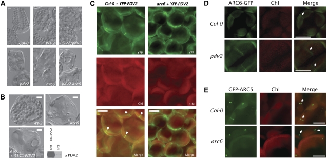
Figure 6.
ARC6 Acts Upstream of PDV2.
(A) Phenotypes observed in the F2 population from arc6-1 × pdv2-1 reciprocal crosses. arc6 pdv2 double mutants were confirmed by PCR-based genotyping and were phenotypically indistinguishable from arc6 mutants.
(B) Phenotypes of mesophyll cells from Wassilewskija-2 (Ws-2; top left), arc6-1 (top right), and arc6-1 transformed with 35Spro-PDV2 and expressing high levels of PDV2 protein (bottom left). Relative PDV2 protein levels in arc6-1 and arc6-1 mutants expressing 35Spro-PDV2 are shown by immunoblot (bottom right).
(C) Localization of YFP-PDV2 in young leaf cells of Col-0 (left panels) and in an arc6 T-DNA mutant (SAIL_693_G04; right panels). Arrowheads indicate midplastid YFP-PDV2 rings.
(D) Localization of ARC6-GFP in young leaf cells of Col-0 (top panels) and pdv2-1 (bottom panels). Arrows indicate midplastid ARC6-GFP rings.
(E) Localization of GFP-ARC5 in young leaf cells of Col-0 (top panels; arrows indicate midplastid GFP-ARC5 localization) and in an arc6-1 mutant (bottom panels; arrows indicate cytosolic GFP-ARC5 patches).
Bars = 10 μM.

